
Spectral Doppler: Tricuspid Valve
Obtaining the spectral Doppler
- Identify the tricuspid valve (TV) in the mid-esophageal views. Each of the following three two-dimensional TEE views can be used:
- Align the pulsed wave (PW) cursor through the TV and place the sample volume box at the TV leaflet tips during diastole (figure 1).
- Note: Adequate Doppler alignment of the TV can be challenging and may require off-axis images.
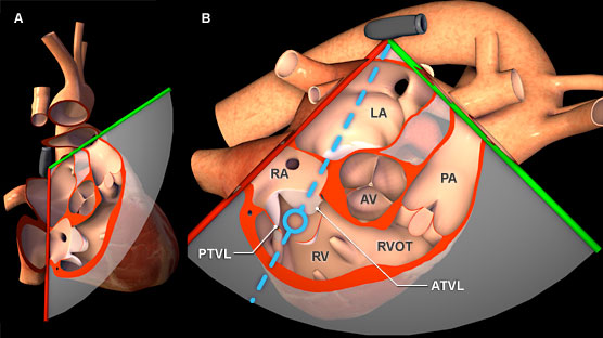
Figure 1: Three-dimensional heart model shown in cross-section to highlight the position of the TEE plane in the mid esophageal right ventricular inflow-outflow view. A) Anterior view of the heart with TEE plane. B) Right lateral view of the heart and TEE plane. The blue dotted line represents the orientation of the pulsed wave cursor and the blue circle indicates the position of the sample box at the tip of the tricuspid valve leaflets. Key: ATVL = Anterior/Septal Tricuspid Valve Leaflet, AV = Aortic Valve, LA = Left Atrium, PTVL = Posterior Tricuspid Valve Leaflet, PV = Pulmonic Valve, PA = Pulmonary Artery, RA = Right Atrium, RVOT = Right Ventricle Outflow Tract.
Features of tricuspid valve spectral Doppler
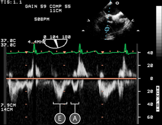
Figure 2: Spectral doppler data acquired for blood flow through the tricuspid valve. In the upper right, a two dimensional TEE image of the mid-esophageal right ventricular outflow view; the blue circle indicates the location of the sample volume at the tip of the tricuspid valve leaflets. In the lower half of the image, a spectral doppler trace shows the relationship between blood velocity and time. The baseline is orange. Key: E = Early diastolic filling wave, A = Atrial kick wave.
- The on screen representation is two peaked with Early diastolic filling (E) and Atrial kick (A) waves seen below the baseline as the direction of blood flow is away from the transducer (figure 2).
- The E wave has Maximum velocity (Emax) of 40 ± 9.8 cm/sec
- The A wave has a Maximum velocity (Amax) of 2.6 ± 7.5 cm/sec
- Blood flow velocity is the lowest through the TV given its large orifice area.
Physiological variation
- Pre/afterload
- Respiratory variation (↑ during spontaneous inspiration, ↓ positive pressure inspiration)
Pathological variation
- Diastolic dysfunction
- Arrhythmias
- Tricuspid valve disease
- Pericardial disease
- Cardiomyopathies