
Spectral Doppler: Pulmonary Veins
Obtaining the spectral Doppler
- Identify the pulmonary veins (PV) in the mid-esophageal views. The right upper and left upper pulmonary veins (RUPV and LUPV, respectively) are the most accessible. The following four two-dimensional TEE views can be used:
- To aid identification of the pulmonary veins, apply colour Doppler using a low velocity scale (< 40 cm/sec).
- To assess flow in a pulmonary vein, place the pulsed wave (PW) sample volume box > 0.5 cm within the centre of the vein (figure 1).
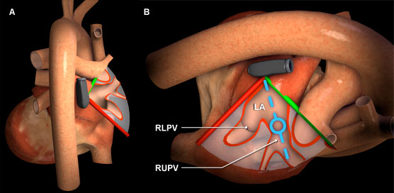
Figure 1: Three-dimensional heart model shown in cross-section to highlight the position of the TEE plane in the modfied right pulmonary vein view. A) Posterior view of the heart with TEE plane. B) Right posterolateral view of the heart and TEE plane .The blue dotted line represents the orientation of the pulsed wave cursor and the blue circle indicates the position of the sample box in the right upper pulmonary vein. Key: DescAor = Descending aorta, LA = Left atrium,RLPV = Right lower pulmonary vein, RUPV = Right upper pulmonary vein.
Features of pulmonary veins spectral Doppler
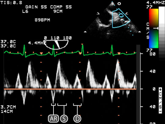
Figure 2: Spectral doppler data acquired for blood flow through the right upper pulmonary vein. In the upper right, a two dimensional TEE image of the modified bicaval view; the blue circle indicates the location of the sample volume which is placed within the right upper pulmonary vein. In the lower half of the image, a spectral doppler trace shows the relationship between red blood cell velocity and time. The baseline is orange. Key: D = Diastolic wave, AR = Atrial systolic reversal and S = Systolic wave.
- A normal pulmonary vein spectral Doppler trace has three waves (figure 2):
- The Systolic (S) wave is caused by antegrade ventricular systolic flow and is seen above the baseline. The velocity (VS) is 30 – 80 cm/sec. The S wave has 2 components most pronounced during increased P-R interval of ECG:
- S1 due to atrial relaxation that is determined by LA pressure, contraction and relaxation.
- S2 due to mitral annular descent and impacted by stroke volume and pulse wave propagation in the pulmonary arterial tree.
- The Diastolic (D) wave is caused by antegrade ventricular diastolic flow and is seen above the baseline. The Velocity (VD) is 20 – 70 cm/sec.
- The Atrial Systolic Reversal (AR) wave caused by retrograde atrial systolic flow, is seen below the baseline. The Velocity (VAR) is 10 – 25 cm/sec. Duration (ARdur) is 30-160 msec.
- The Systolic (S) wave is caused by antegrade ventricular systolic flow and is seen above the baseline. The velocity (VS) is 30 – 80 cm/sec. The S wave has 2 components most pronounced during increased P-R interval of ECG:
- Under normal conditions, S ≥ D.
- Pulmonary vein ARdur minus the transmitral A wave < 25 msec.
Physiological variation
- Aging (↑ S/ D, ↑ VAR)
- Tachycardia causes ↑ S and ↓ D
Pathological variation
- Diastolic dysfunction
- Mitral valve disease
- Arrhythmias
- Pericardial disease