
Biomedical Communications Masters Research Projects
Biomedical Communications (University of Toronto) offers a two year M.Sc.BMC program with training in medical illustration, 3D modeling, animation, user-centered design, and interactive educational application development. The PIE team application developers are all graduates of this program, and we continue to work with current students in developing projects for fulfillment of the requirements for their Masters degree.
VIRTUAL Liver
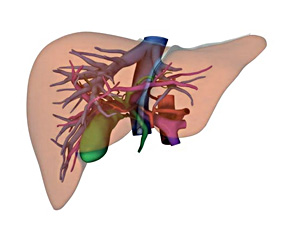
This aim of this project was to build an online tool to help students learn the surgical anatomy of the liver. The project involved the creation of an online application which featured an interactive 3D model of the liver. The tool allowed users spin the liver in two planes and view each of the internal structures selectively. Click here to read more about the current state of the project.
Student:
Supervisor:
Professor Jodie Jenkinson, University of Toronto, Biomedical Communications
Clinical content experts:
Dr. Carol-anne Moulton, University of Toronto Department of Surgery
Dr. Steve Gallinger, University of Toronto Department of Surgery
VIRTUAL bronchoscopy
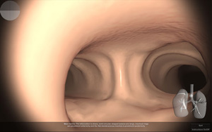
The aim of this project was to develop an online virtual bronchoscopy exam. Application development involved interface design, application coding and the creation of an anatomically accurate 3D model of the bronchial tree. The latter was built using Autodesk Maya and then imported into the gaming engine.
Student:
Supervisor
Professor Marc Dryer, University of Toronto, Biomedical Communications
Clinical content experts:
Dr. Adriaan Van Rensburg, Toronto General Hospital Department of Anesthesia
Dr. George Kanelakos, Toronto General Hospital Department of Anesthesia
VIRTUAL Nerve Block
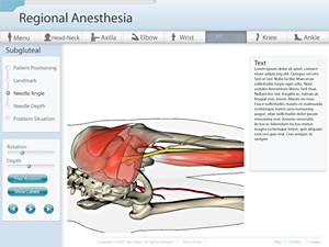
The aim of this project was to create an online tool that could help teach anesthesiolgists how to perform peripheral nerve blocks. An anatomically correct three-dimensional model of the hip with bones, muscles, arteries, veins and nerves was built. An interface, employing interactively adjustable viewpoints, was designed that allowed users to explore the relative positions of needle and tissues.
Student:
Supervisor:
Professor Jodie Jenkinson, University of Toronto, Biomedical Communications
Professor Nicholas Woolridge, University of Toronto, Biomedical Communications
Clinical content expert:
Dr. Marcin Wasowicz, Toronto General Hospital Department of Anesthesia
Axillary Lymph Node Dissection
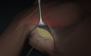
The purpose of this project was to create a web-based learning tool for surgical residents to understand the ALND procedure. Although the surgery is slowly being replaced with the sentinel lymph node biopsy (SLNB) procedure, it is still necessary in treating patients with advanced breast cancer. As ALND cases decline, it becomes more difficult for surgical residents to learn the procedure by direct observation. Therefore, the aim was to create a short 3D animation that depicts the general flow of the surgery, highlighting important anatomical features and 'pearls' of knowledge.
Student:
Supervisor:
Professor Jodie Jenkinson, University of Toronto, Biomedical Communications
Clinical content expert:
Dr. Tulin Cil, Department of Surgical Oncology, Princess Margaret Hospital