
Cardiac embryology
The need
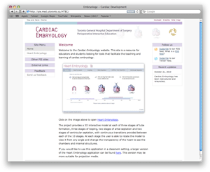
Click on the image to open the Cardiac Embryology module in a new window.
Explaining the processes underlying the development of the heart is one of the most demanding tasks in teaching embryology. There are three key aspects of this development that present challenges:
- The complex transformations in the exterior shape of the organ.
- The formation of the interior chambers and great vessels.
- The origin and migration of the cells that form the heart.
The problem
Current visual aids for teaching cardiac embryology consist for the most part of two-dimensional drawings or scanning electron microscopy images at a number of fixed stages of the developing heart. These illustrations do not capture the complex process of transformation between the stages. It is also difficult to visualize the convoluted shape of the exterior of the heart and the formation of the interior chambers.
The solution
The project will provide a 3D interactive model at each of three stages of tube formation, three stages of looping, two stages of artial septation and two stages of ventricular septation, with continuous transitions provided between each of the 10 stages. At each stage the user will be able to rotate the model to view it from any angle and change the transparency of the heart to see the chambers and internal structures.