
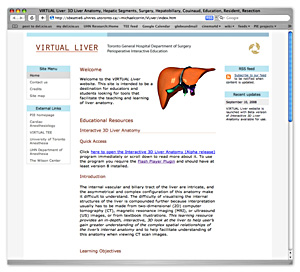
VIRTUAL Liver website
Introduction
Click on the image to open the Virtual Liver website in a new window.
The VIRTUAL Liver site is intended to be a resource for educators and students looking for tools that facilitate the teaching and learning of liver anatomy. The site is a collaborative effort of the PIE group (Toronto General Hosiptial Department of Anesthesia and Pain Management) and the University of Toronto Department of Surgery.
Click here to open the VIRTUAL Liver site in a new window.
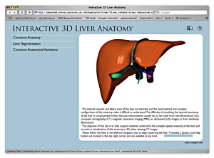
Interactive 3D Liver Anatomy Module
Click on the image to open the Virtual Liver website in a new window.
The internal vascular and biliary tract of the liver are intricate, and the asymmetrical and complex configuration of this anatomy make it difficult to understand. The difficulty of visualising the internal structures of the liver is compounded further because interpretation usually has to be made from two-dimensional (2D) computer tomography (CT), magnetic resonance imaging (MRI), or ultrasound (US) images, or from textbook illustrations. This learning resource provides an in-depth, interactive, 3D look at the liver to help user’s gain greater understanding of the complex spatial relationships of the liver’s internal anatomy and to help facilitate understanding of this anatomy when viewing CT scan images.