Content
Lung Ultrasound
![]() 2020-12-31: Lung Ultrasound is now available in HTML5
2020-12-31: Lung Ultrasound is now available in HTML5
This module has been converted from Adobe Flash to HTML5, and is now accessible on tablets and smartphones as well as on desktop computers. At this time, only the core module with the normal views has been converted to HTML5. The views with pathology (pneumothorax and pleural effusion) are in the process of being converted and will be available early in the new year (2021).
Introduction
Lung ultrasound has been successfuly used in diagnosing the cause of respiratory failure in the Emergency Department and has been shown to have higher sensitivity and specificity than chest X ray and physical examination in detecting pleural effusions and pneumothorax. Point of Care Lung Ultrasound is gaining popularity across many different medical specialties and is becoming part of the modern physicians clinical armamentarium.
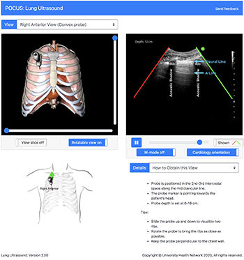
We have created this online interactive module to assist with teaching and learning the use of ultrasound in the assessment of the lung. Users can view the ultrasound recordings for each of 6 probe positions and see a corresponding 3D model of the probe, ultrasound plane, and torso for each view. Different types of probes may be selected that are appropriate for the view. The user can choose B-mode and M-mode imaging modalities for the anterior views. Lung US is mostly based on artifacts, and this module will provide explanations for these artifacts in the normal lung. The module will be expanded in the coming months to include examples of common lung pathologies.
How to Use the Module
Each lung ultrasound view can be selected by clicking on a probe position indicated by the dotted outline of probes on the model in the lower left panel. Different probe types may be selected from buttons at the bottom of this panel.
For each view, the 3D model of the probe, ultrasound plane, lung, rib cage and diaphragm can be rotated in the horizontal or vertical plane to view it from any angle. The entire anatomical model can be cut along the ultrasound plane and rotated. The model can then be oriented so the structures correspond to the ultrasound image.
Acknowledgements
The development of this module was supported by the University of Toronto OIME Summer EIT Program and a grant from the Academic Health Science Centres Academic Funding Plan Innovation Fund. The Lung Ultrasound module was developed by Jean YiChun Lin, MScBMC with medical content provided by Dr. Massimiliano Meineri, Dr. Alberto Goffi and Dr. Catherine Nix, and with consultation from Dr. Gordon Tait.